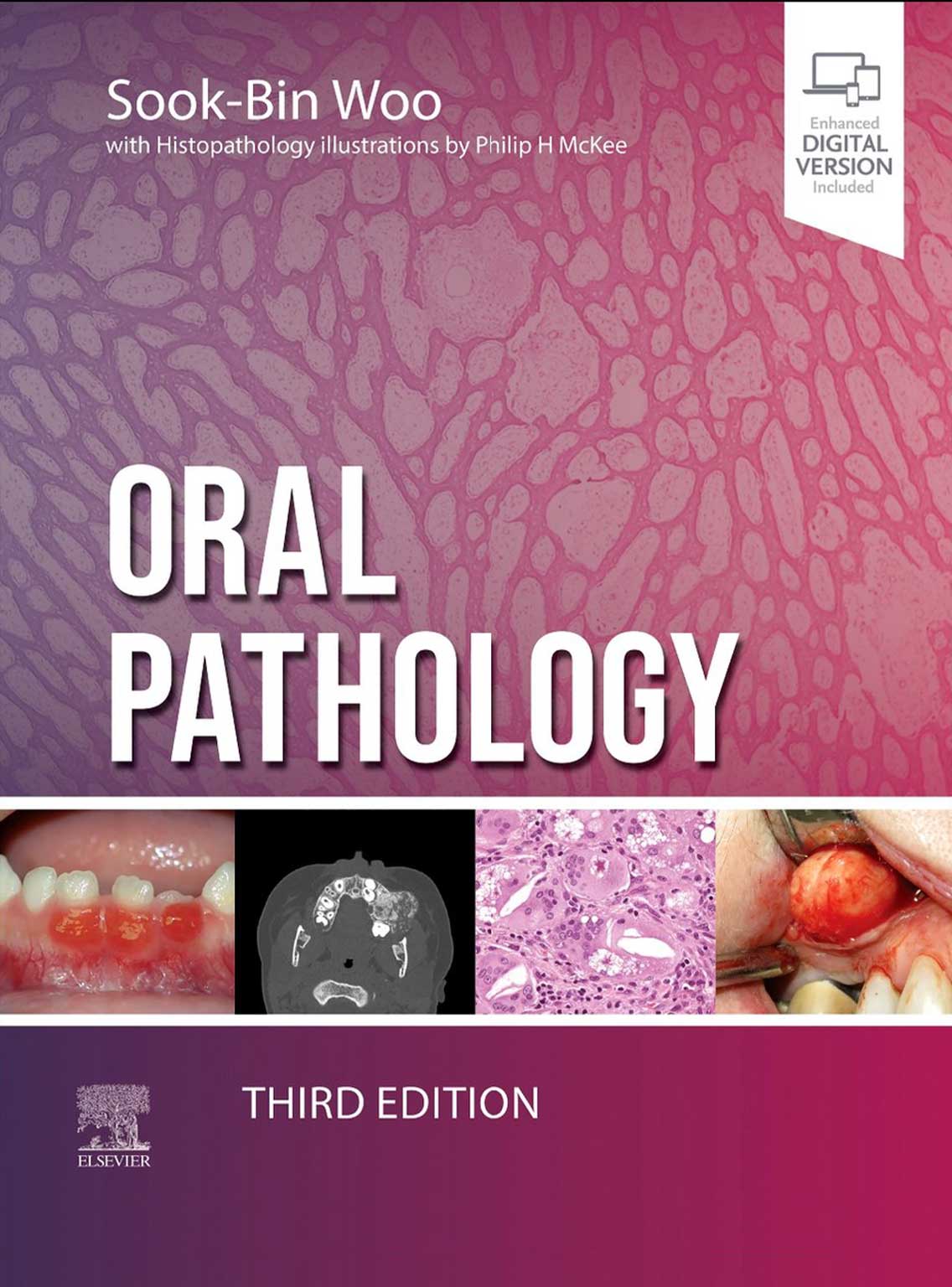
Oral Pathology 2024 (Third Edition)
۱۸,۸۰۰,۰۰۰ ریال قیمت اصلی: ۱۸,۸۰۰,۰۰۰ ریال بود.۱۵,۹۸۰,۰۰۰ ریالقیمت فعلی: ۱۵,۹۸۰,۰۰۰ ریال.
| Author | |
|---|---|
| Edition Number | |
| Copyright Year | |
| Print color | |
| Page Count | |
| Cover Type | |
| Dimensions | |
| Paper type | |
| Publishers | |
| ISBN Number | |
| Weight |
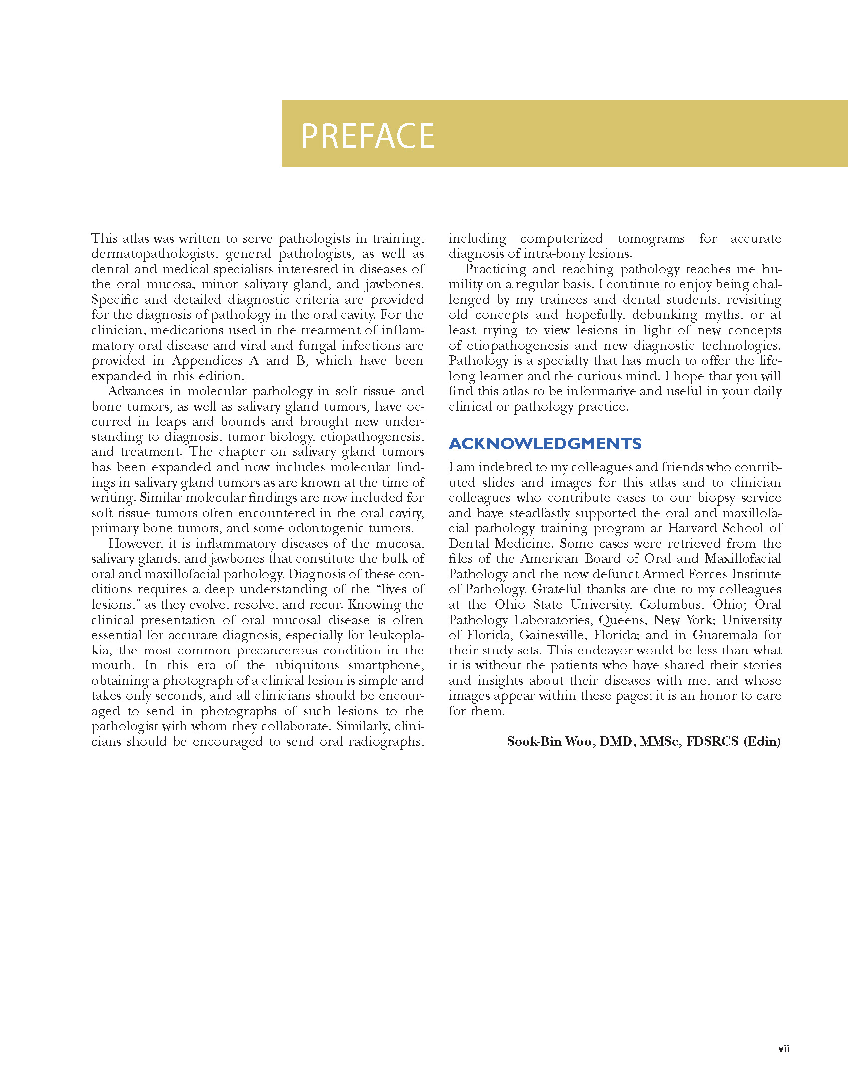
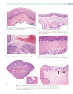
This atlas was written to serve pathologists in training, dermatopathologists, general pathologists, as well as dental and medical specialists interested in diseases of the oral mucosa, minor salivary gland, and jawbones. Specific and detailed diagnostic criteria are provided for the diagnosis of pathology in the oral cavity. For the clinician, medications used in the treatment of inflammatory oral disease and viral and fungal infections are provided in Appendices A and B, which have been expanded in this edition. Advances in molecular pathology in soft tissue and bone tumors, as well as salivary gland tumors, have occurred in leaps and bounds and brought new understanding to diagnosis, tumor biology, etiopathogenesis, and treatment. The chapter on salivary gland tumors has been expanded and now includes molecular findings in salivary gland tumors as are known at the time of writing. Similar molecular findings are now included for soft tissue tumors often encountered in the oral cavity, primary bone tumors, and some odontogenic tumors. However, it is inflammatory diseases of the mucosa, salivary glands, and jawbones that constitute the bulk of oral and maxillofacial pathology. Diagnosis of these conditions requires a deep understanding of the “lives of lesions,” as they evolve, resolve, and recur. Knowing the clinical presentation of oral mucosal disease is often essential for accurate diagnosis, especially for leukoplakia, the most common precancerous condition in the mouth. In this era of the ubiquitous smartphone, obtaining a photograph of a clinical lesion is simple and takes only seconds, and all clinicians should be encouraged to send in photographs of such lesions to the pathologist with whom they collaborate. Similarly, clinicians should be encouraged to send oral radiographs, including computerized tomograms for accurate diagnosis of intra-bony lesions. Practicing and teaching pathology teaches me humility on a regular basis. I continue to enjoy being challenged by my trainees and dental students, revisiting old concepts and hopefully, debunking myths, or at least trying to view lesions in light of new concepts of etiopathogenesis and new diagnostic technologies. Pathology is a specialty that has much to offer the lifelong learner and the curious mind. I hope that you will find this atlas to be informative and useful in your daily clinical or pathology practice.
ACKNOWLEDGMENTS I am indebted to my colleagues and friends who contributed slides and images for this atlas and to clinician colleagues who contribute cases to our biopsy service and have steadfastly supported the oral and maxillofacial pathology training program at Harvard School of Dental Medicine. Some cases were retrieved from the files of the American Board of Oral and Maxillofacial Pathology and the now defunct Armed Forces Institute of Pathology. Grateful thanks are due to my colleagues at the Ohio State University, Columbus, Ohio; Oral Pathology Laboratories, Queens, New York; University of Florida, Gainesville, Florida; and in Guatemala for their study sets. This endeavor would be less than what it is without the patients who have shared their stories and insights about their diseases with me, and whose images appear within these pages; it is an honor to care for them. Sook-Bin Woo, DMD, MMSc, FDSRCS (Edin)
